Highlights of our Work

2024 | 2023 | 2022 | 2021 | 2020 | 2019 | 2018 | 2017 | 2016 | 2015 | 2014 | 2013 | 2012 | 2011 | 2010 | 2009 | 2008 | 2007 | 2006 | 2005 | 2004 | 2003 | 2002 | 2001
DNA graphene

image size: 210.7KB
made with VMD

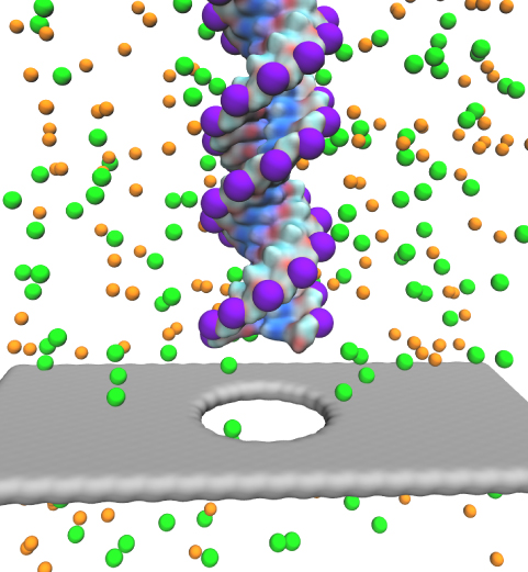
Characterizing the genetic makeup of individuals can help select the best treatment for individuals, but requires that sequencing of the whole DNA of patients can be achieved for less than $1000. Fortunately, recent discoveries in physics promise help. Indeed, the discovery of the thinnest material known to mankind, graphene, promises a new and cheaper way to sequence human DNA. As reported in the December 2011 highlight, graphene pores can conduct electrophoretically DNA through very small pores, so-called nanopores. A new study has demonstrated that graphene, forming a single atomic layer thin stripe with a nanopore in the middle, can conduct an electrical current, the sheet current, around the pore. The sheet current is sensitive to the DNA passing through the pore and may even sense the passing DNA's sequence. In this case monitoring the current can establish a DNA sequence reader. The sensitivity of the sheet current depends critically on an orderly passage of DNA. Optimal sensitivity can arise when the passing DNA is stretched mechanically. Molecular dynamics simulations using NAMD suggest conditions for mechanically manipulating DNA for optimal sequence analysis. More on our graphene nanopore website.
Integrin-RGD binding under force

image size: 507.8KB
made with VMD

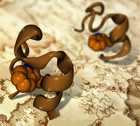
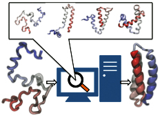
Every protein in a living cell needs to assume a proper structure in order to fulfill its biological function. The proteins that fails to do so cause malfunction of cells, often leading to disastrous diseases. Thus, how a protein folds into its native structure is one of the central biological processes that need to be understood. The modern view of protein folding suggests that a folding process is governed by structural transitions among a wide spectrum of structures that a protein could access; all of these possible structures and associated transitions constitute a complex roadmap for protein to follow, very much like the one commonly used to navigate a car to its destination through the best route. Obtaining comprehensive and accurate roadmaps for protein folding are essential for the characterization of correct folding routes. In theory, such a roadmap could be explored through computational simulations using an accurate model including every atomistic detail; in practice, the structural complexity of proteins turns the exploration of its roadmap into a daunting computational task. As reported recently, researchers have demonstrated an efficient way to explore roadmaps for protein folding by utilizing a hybrid model which combines atomic models for the protein part with a fast simplified model for the surrounding water part in order to achieve both accuracy and efficiency. By investigating folding mechanisms of two proteins, the researchers showed that their approach is able to generate folding roadmaps as accurate as those obtained with complete atomic models while only taking days to finish the task, much faster than the complete atomic models that usually need months. For details, please see our hybrid-resolution model website.
Ion Diffusion on a Grid near MscS

image size: 411.0KB

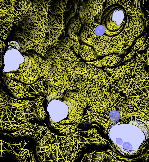
Traffic flow in a city is affected by large-scale features, like the layout of road networks, as much as it is by small-scale ones, like traffic lights at a road junction. Likewise, the transport of small molecules in cells occur on multiple scales. For example, ions diffusing through the mechanosensitive channel of small conductance (MscS) (see highlights from Jul 2011, "Smart Bacterial Safety Valve", Mar 2008, "Observation and Simulation depict Cell's Safety Valve", Feb 2007, "Observing and Modeling a crucial Membrane Channel", May 2006, "Electrical Safety Valve", and Nov 2004, "Japanese Lantern Protein") must navigate the intricate geometry of the MscS, which varies by the Ångstrom. At the same time, the distribution of ions within hundreds of Ångstroms of the MscS fluctuate as ions escape through the channel, thus changing the electrostatic landscape seen by other ions as they approach the MscS. In order to model both the fine and bulk aspects of diffusion in systems like that of the MscS, scientists have proposed, in a recent report, a method that marries the high spatial resolution of molecular dynamics to long range diffusion. In the new description, biomolecules "diffuse" by hopping through a grid under the influence of Coulomb and other forces. More on our Kinetic Diffusion web site.
The Force Field Toolkit

image size: 307.6KB

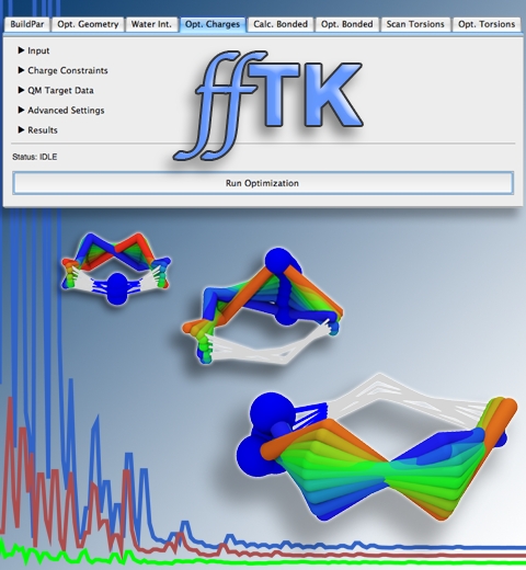
Structural biologists are increasingly turning to simulation methods to investigate the connections between molecular structure and biological function. Classical molecular dynamics (MD) simulations, such as those performed by the simulation software NAMD, rely on potential energy functions requiring parameters to describe atomic interactions within the molecular system. While these parameters are available for the most commonly simulated biopolymers (e.g., proteins, nucleic acids, carbohydrates), many small molecules and other chemical species lack adequate descriptions. The complexity of developing these parameters severly restricts the application of MD technologies across many fields, including most notably drug discovery. Recently, researchers have developed software, the Force Field Toolkit (ffTK), that greatly reduces these limitations by facilitating the development of parameters directly from first principles. ffTK, distributed as a plugin for the molecular modeling softare VMD, addresses both theoretical and practical aspects of parameterization by automating tedious and error-prone steps, performing multidimensional optimizations, and providing quantitative assessment of parameter performance--all from within an easy-to-use graphical user interface. Additional information on ffTK, including documentation and screencast tutorials, can be found here.
Quantum biology

image size: 679.7KB
credit: Olga Svinarski

Clearly, the laws of physics hold and are exploited in living organisms. Speaking as a physicist, most biological characteristics stem from the laws of classical physics that students learn in their first year. However, crucial characteristics in organisms are governed by quantum physics, a higher and still active area of physics. The latter characteristics are those in which biological processes involve the jumps of electrons from one state to another state: electrons are exemplary quantum particles. The quantum behavior of electrons cover all chemical transformations, for example in case of formation or breaking of chemical bonds, but it arises also in optical transitions induced through light absorption by biomolecules. Quantum behavior of electrons in such cases is localized in single molecules, but it can also spread over many biomolecules in a typical quantum mechanical fashion. The biomolecules form in such case a chorus that sings in one coherent voice, rather than chatters incoherently. This type of behavior is not only of interest to the modern biological researcher, but also to modern physics researchers working on quantum computing. Three recent reviews summarize fascinating quantum behavior in biology as it comes about in vision, photosynthesis, and animal navigation using the earth magnetic field. In case of vision (see July 2003, March 2002 and August 2001 highlights) twelve electrons in a molecule called retinal correlate their motion during light absorption and steer the optically excited molecule to alter its shape, thereby initiating an extremely sensitive response to light. In case of photosynthesis (see April 2010, August 2010, March 2002 and September 2001 highlights), molecules of chlorophylls utilize thermal effects to optimally absorb sun light and then share the resulting electronic excitations among themselves through quantum coherence. In case of animal navigation (see February 2012, July 2010, July 2009, and April 2007 highlights), quantum effects apparently bring about a magnetic compass that can sense the Earth field through its interactions of biomolecules despite the fact the interaction energy amounts to only a tiny fraction of thermal energy present at body temperature; physicists are eager to learn the trick as it might teach them how to build quantum computers without costly cooling to extremely low temperatures. More on vision here, here, and here, on photosynthesis here, here and on animal navigation here.
RHDV

image size: 651.0KB
made with VMD

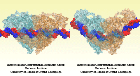
DNA, a long linear molecule, is the carrier of genetic information. In the cell, each DNA molecule is packaged in a structure called chromosome. The ends of linear chromosomes are capped by structures known as telomeres to prevent fusion with neighboring chromosomes. Telomeres are maintained by an enzyme called telomerase during DNA replication. In order to do so, telomerase has to find the telomere region on DNA quickly and precisely. One telomerase is the protelomerase TelK, which binds to the ends of DNA, cleaves DNA strands and refolds cleaved DNA ends into hairpin telomeres in linear chromosomes of prokaryotes and viruses. Previous studies have shown that TelK is only active as a dimer. In a recent study, researchers investigated the target-search mechanism of protelomerase TelK through single-molecule experiments and molecular dynamics simulation. It was revealed that as a monomer, TelK undergoes one-dimensional diffusion along non-specific DNA (without telomere sequence), and is able to bind to the target site preferentially. There, the target-immobilized monomer waits for a second binding partner to form an active protein complex. More on our TelK website.
HIV-1 Capsid

image size: 467.0KB
see also movie: 15.5MB
made with VMD

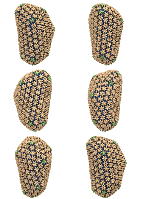
Human immunodeficiency virus type 1 (HIV-1) is the major cause of AIDS, for which treatments need to be developed continuously as the virus becomes quickly resistant to new drugs. When the virus infects a human cell it releases into the cell its capsid, a closed, stable container protecting the viral genetic material. However, interaction with the cell triggers at some point an instability of the capsid, leading to a well timed release of the genetic material that merges then with the cell's genes and begins to control the cell. The dual role of the capsid, to be functionally both stable and unstable, makes it in principle an ideal target for antiviral drugs and, in fact, treatments of other viral infections successfully target the respective capsids. The size of the HIV-1 capsid (about 1,300 proteins), and its irregular shape had prevented so far the resolution of a full capsid atomic-level structure. However, in a tour de force effort, groups of experimental and computational scientists have now resolved the capsid's chemical structure (deposited to the protein data bank under the accession codes 3J3Q and 3J3Y). As reported recently (see also journal cover), the researchers combined NMR structure analysis, electron microscopy and data-guided molecular dynamics simulations utilizing VMD to prepare and analyze simulations performed using NAMD on one of the most powerful computers worldwide, Blue Waters, to obtain and characterize the HIV-1 capsid. The discovery can guide now the design of novel drugs for enhanced antiviral therapy. More information is available on our virus website, in video, and in a press release.
LOV domain

image size: 1.9MB
made with VMD

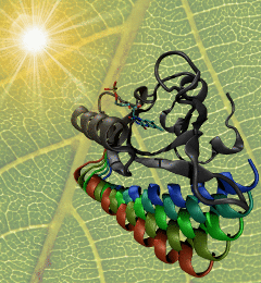
The ability to sense light is crucial for both plants and animals; animals use their vision to navigate and interact with their surroundings, whereas plants grow toward light to optimize photosynthesis. One of the most important photosensors in plants relies on tiny molecular switches known as LOV domains. When light strikes a LOV domain, it causes the formation of a single chemical bond; the unique structure of the LOV domain converts this subtle change into a protein unfolding event that triggers signaling. The mechanism through which LOV domains amplify bond formation into large-scale molecular motion is of great interest both for designing light-activated proteins for synthetic biology applications, and as a model for understanding the harder-to-study molecular switches that govern most of the functions of living cells. As reported recently, researchers used a series of long-timescale molecular dynamics simulations to show the locations of molecular levers that allow light-induced bond formation to rearrange the entire structure of the LOV domain. The simulations highlighted two main paths of information flow from the heart of the photoreceptor to the surface of the protein, giving unprecedented insight into the function of this light-activated molecular switch. More information is available on the biological photoreceptors website.
RHDV

image size: 788.0KB
see also movie, 6.4MB
made with VMD

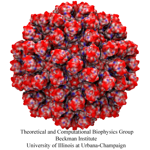
Rabbit hemorrhagic disease is extremely contagious and associated with liver necrosis, hemorrhaging, and high mortality in adult rabbits. First described in China in 1984, within a few years, rabbit hemorrhagic disease spread to large parts of the world and today threatens the rabbit industry and related ecology. The disease is caused by a virus, aptly named rabbit hemorrhagic disease virus. As reported recently, a group of experimental and computational researchers combining crystallography, electron microscopy and data-guided molecular dynamics simulations utilizing NAMD determined an atomic model of the capsid, namely the protein shell that surrounds the genetic material of the virus. The capsid simulations involved 10 million atoms and have become feasible only through Blue Waters, a brand new petascale supercomputer. The atomic model, analyzed by means of VMD, recently adapted to studies of very large structures, resolves the structural framework that furnishes both mechanical protection to the viral genes as well as a quick release mechanism after a virus enters a host cell. Researchers can use the detailed knowledge of the capsid structure to develop vaccines against rabbit hemorrhagic disease. More information is available on our virus website and in a news story.
SecY

image size: 223.2KB
made with VMD

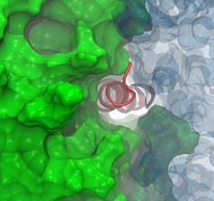
For newly made membrane proteins, getting to their final destination in the membrane requires another protein, the channel SecY, to provide a pathway (see the Sept. 2007 and Feb. 2011 highlights). But just knowing the route is not enough, because SecY presents the nascent protein with a choice: insert into the membrane or cross the channel to the watery exterior. How the nascent protein comes to a decision has long been a point of uncertainty, although it has been presumed to be driven by purely energetic considerations, i.e., the protein goes to the environment it ultimately prefers. Now, recent simulations and free-energy calculations spanning time scales from nanoseconds all the way to seconds have revealed that how long the nascent protein deliberates in the channel is just as great a factor in its final location as how favorable it is there. It was found that the longer the protein takes to decide, the more likely it is to choose the membrane, proving that, at least for membrane insertion, slow and steady wins out. More information can be found on the protein translocation website.
PHI: Parallel Hierarchy Integrator

image size: 1005.1KB

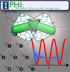
Quantum mechanics rules all natural processes, but is manifested most strongly when acting on the lightest particles, namely the well-known electrons. To study quantum effects physicists routinely resort to very low temperature, that of liquid helium. Amazingly, living systems seem to exploit quantum effects for their benefit, but do so at temperatures typical for life, namely around room temperature or warmer. A particularly important case is photosynthetic light harvesting where so-called quantum coherence plays a critical role when electrons in assemblies of chlorophylls become excited by sun light and the excitation energy is harvested by utilizing it to charge photosynthetic membranes. In order to understand how photosynthesis can exploit room temperature quantum effects one needs to know how the temperatures, which are much higher than those in the physics laboratories where liquid helium is employed for cooling, affect electron behavior. The knowledge can be gained by so-called dissipative quantum mechanical descriptions, but the needed computer calculations are extremely demanding. To address this demand, researchers have developed the software PHI that uses the power of parallel computers, as described in a recent report. PHI has already been used to understand how many chlorophyll molecules act together to absorb sunlight among themselves and let the excitation migrate between chlorophylls to so-called reaction centers where the excitation energy is converted into a membrane potential. The overall light harvesting process has been described in various reports (1, 2, 3) and in a review (4). The PHI software can be obtained from our web site. More information on PHI is available here.
BAR domain

image size: 1.3MB
Olga Svinarski and VMD

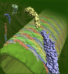
The cells of higher life forms, so-called eukaryotic cells, are subdivided through many internal membranes made of lipid bilayers. The internal membranes assume numerous shapes, like spheres, tubes or parallel sheets. Outside of cells, biological membranes adopt usually flat shapes and the question arises, how do eukaryotic cells sculpt their inner membranes? The question of flat membrane sculpting is particularly interesting also as mature cells constantly produce new membrane shapes, for example spherical vesicles filled with certain biomolecules destined for release into the extracellular space, a process called exocytosis. The cell has many mechanisms available for sculpting its membranes, one of them relying on proteins called BAR domains that act from the surface of lipid bilayers. Molecular modeling with NAMD and VMD has provided valuable views of BAR domains at work in case of the so-called N-BAR family (see the earlier highlights Protein Teamwork, Jun 2009 and Proteins Sculpting Cell Interior, Sep 2008). Researchers report now an extension of the earlier studies to the F-BAR domain family of membrane sculpting proteins. The new modeling work is particularly exciting as it can be directly compared to electron microscopy images of membrane tubes sculpted from flat membranes in experiments done outside of cells. The new studies reveal how F-BAR domains sculpt tubular membranes through the shape of dimerized domains and through F-BAR domains not acting individually, but as an army of F-BAR domains adopting an ordered formation on one side of the membrane. More on our F-BAR domain web page.
Integrin-RGD binding under force

image size: 64.7KB
see also movie, 11.5MB
made with VMD

Every living cell relies on proteins to carry out its functional tasks; every protein needs to assume a proper shape in order to be operational for these tasks. How a protein, composed of a particular sequence of amino acids, could find its way to a proper shape is a fundamental, yet mysterious biological process. Researchers have sought to unravel atomistic details of protein folding processes through computer simulations, but modeling such processes is computationally demanding. It was only recently that some researchers have been able to observe in some case how proteins fold, but needed for the purpose the fastest computers available today. One of these computers is Anton, the expensive special-purpose supercomputer available essentially only to a single research group. Is there an affordable way to simulate protein folding? One solution could be coarse-grained methods. These methods save tremendous computational effort by replacing computational models that include all atomistic detail. However, the simplified models need to include a sufficiently accurate description of proteins for modeling folding processes. As reported recently, researchers have overcome the challenge by combining atomistic and coarse-grained descriptions. The new method is fast enough to follow movements of proteins long enough to see them fold, while requiring only readily available computer powers. The new method allowed researchers to analyze complete folding events for seven proteins, including a protein, called α3D (see movie, 11.5 M), that is one of the largest proteins ever folded computationally. More on our hybrid-resolution model website.
Electron transfer in cryptochrome

image size: 90.9KB

Animals and plants, together with other life forms, possess internal clocks that attune them to the daily rhythm on Earth. A sign of such clocks is jet lag, the discomfort experienced by humans when due to travel across several time zones our internal clocks need to be reset to the new time zone. Feed-back to local day light assists the resetting and a key light receptor serving the purpose is a protein called cryptochrome. The name was chosen as the receptor hid for a long time from the instruments of researchers, but today the name seems still appropriate as the physical mechanism of the receptor is shrouded in mystery and subject to dispute. Adding to the mystery is an apparent second role of cryptochrome, namely that of a sensor for the Earth' magnetic field, which helps migratory birds and many other animals in long-range navigation (see our magnetoreception page). The biological function of cryptochrome supposedly arises from a photoactivation reaction involving electron transfer, but the reaction pathway is difficult to resolve experimentally as the best available method, time-resolved spectroscopy, cannot identify unequivocally the photoproducts produced through cryptochrome light absorption. Experimentalists hate to admit the calamity, but likely the only way out are a combination of quantum-chemical and classical molecular dynamics calculations. Such calculations were recently performed and the results reported. The calculations demonstrate that after absorption an electron is transferred inside cryptochrome, the new state becomes stabilized through proton transfer and decays back to the protein's resting state on time scales allowing the protein, in principle, to act as a light as well as magnetic sensor. More details can be found on our cryptochrome webpage.

2024 | 2023 | 2022 | 2021 | 2020 | 2019 | 2018 | 2017 | 2016 | 2015 | 2014 | 2013 | 2012 | 2011 | 2010 | 2009 | 2008 | 2007 | 2006 | 2005 | 2004 | 2003 | 2002 | 2001